- BioPharm International-08-01-2013
- Volume 26
- Issue 8
Clearing Viral Concerns in Animal-Derived Biomaterials
Viruses in animal-derived starting materials could contaminate biopharmaceutical final product. A rigorous testing strategy and removal methods are reviewed.
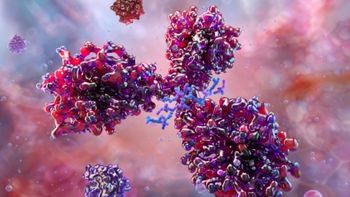
There is a possibility that viruses could be present in animal-derived starting materials used in biopharmaceutical production and, therefore, potentially contaminate the final product. Because of the potential risk to the end user, it is crucial to approach the safety of these therapeutic products through a rigorous and systematic testing strategy. Kate Smith, principal scientist, development services at BioReliance offers insights on the tests required to check for the presence of these contaminants and how to remove the contaminants that are present in final products using chemical inactivation or processes such as chromatography and nanofiltration.
Kate Smith
Material Risks
Biopharmaceutical manufacturing processes frequently rely on starting materials derived from mammalian sources. The level of risks that these materials present are contingent upon the nature of the starting material, mainly, whether they come from human or animal sources.
One example of human-derived material is plasma. Despite the screening of blood donations, when a product is being manufactured from human plasma, there is potential for contamination by pathogens such as hepatitis and human immunodeficiency virus (HIV).
Additionally, many biological drugs are manufactured in Chinese hamster ovary (CHO) or murine cell lines. With these cell lines, it is rare for viruses to be absent. While these materials are well characterized and screened for viruses in advance, the cells commonly contain endogenous viral contamination, typically in the form of a retrovirus. This virus is detected and quantified using transmission electron microscopy as part of the bulk-harvest testing.
This risk is not limited to biopharmaceutical materials. Even products falling into the medical-device category can be affected, such as collagen preparations. Collagen is derived from animal tissues; therefore, there is a high probability that a viral pathogen may be present.
Viruses may also enter the process or product by adventitious means. For instance, a contaminated raw material may be introduced into the process, or viral contamination could have entered the cell culture from a human operator.
The Impacts of Contamination
Historically, on a number of occasions, viral contamination has been detected in products. Perhaps the most well-known case was the vesivirus 2117 contamination discovered in Genzyme's manufacturing facility in Allston, Boston in 2009. As a result, the manufacturing facility was closed for months, causing a supply shortage of the drugs, Cerezyme (used to treat Gaucher's disease) and Fabrazyme (for the treatment Fabry's disease). The incident caused significant problems for patients who needed these medicines.
It was not the first time Genzyme had detected vesivirus in its manufacturing plants. Contamination had also been found at its site in Geel, Belgium. Such incidents highlight the importance of detecting and removing viral contamination at the outset for patient safety. Although patient safety is the greatest concern, these events can also have an effect on business. For example, Genzyme's share prices fell following news of the contamination events, which then became the major factor driving Sanofi-Aventis' acquisition of Genzyme in 2011 (1).
Testing for Viruses
Failing to screen for contamination when working with mammalian starting materials is unacceptable. Viral contamination events can be managed by a number of different approaches, but the first step should always be testing. In general, high-risk materials of biological origin undergo viral clearance, also known as inactivation techniques, to ensure safety. This step is designed to reduce the potential for viral contamination to be introduced into the process from materials in the early stages.
There are regulations regarding testing procedures that must be complied with for biological materials, such as cell stocks and raw materials, used in biopharmaceutical manufacturing. The characterization of master and working cell banks may typically take approximately three months. These tests are completed under strictly regulated guidelines. Testing of cell substrates include identity tests (phenotypic or genotypic characterization), purity tests (free from adventitious microbial agents and cellular contaminants, bacteria, fungi, mycoplasma, and virus), cell-line stability testing and karyology, as well as tumorigenicity.
In-vitro testing is carried out using cell-based assays to screen for potential viral contamination. It is generally done on multiple cell lines that are known to be susceptible to a wide range of human and animal viruses. Three or four different cell lines are typically used, including the species of origin (e.g., CHO) and one susceptible to human viruses. Ultimately, the choice of cell lines will be determined by the viruses likely to be present, whether as an endogenous contamination or as a result of handling of the cells.
In-vivo testing is used to assess for potential viral contamination with those viruses that are unable to grow in in-vitro cell-culture methods. The animal species selected for these studies is defined by the nature and source of the cell substrate being used for the production process. The use of in-vivo studies is carefully controlled, from both regulatory and ethical standpoints.
In addition, other tests that can be used include checking for the presence of retrovirus using electron microscopy or molecular biology techniques such as reverse transcriptase assays. Antibody-production tests are also used to determine if species-specific viruses are present when rodent cell lines are used.
For blood and plasma-derived blood products, the process is slightly different as manufacturers have to rely on donor-screening programs. Various control elements are in place; for example, plasma from donors cannot be fractionated and used in therapeutic products, such as those used to treat hemophiliacs, because of the concerns that remain around bovine spongiform encephalitis (BSE)contamination.
The preferred approach with respect to animal-derived materials is to remove these components from the production process where possible. Significant progress has been made in the replacement of animal-derived materials with recombinant products and plant-derived material. However, even plant-derived raw materials are not without risk.
A new technique called massively parallel sequencing is a powerful tool for detailed, molecular characterization of raw materials. The technology, also known as next-generation sequencing, captures the sequences of millions of nucleic-acid strands in a sample, whether expected or contaminant. While it is not typically used as a routine quality-control (QC) test, it does facilitate an understanding of the universe of potential contaminants. With this additional insight, specific polymerase chain reaction (PCR) based assays can be used as routine QC tests to control the quality of the raw material.
Techniques for Viral Inactivation
Testing and screening of cell substrates and raw materials represents one part of the approach to demonstrate viral safety. The testing strategy will only detect viruses targeted in the assays, and there is the possibility that unknown viruses will remain, for which the assays are not designed to detect. It is, therefore, important to incorporate specific steps into the purification process that will remove or inactivate viruses with different physicochemical properties.
Products such as monoclonal antibodies (mAbs) are typically manufactured using a generic platform type of process. Two key steps are incorporated into the purification of a mAb to prevent viral contamination: incubation at low pH to inactivate enveloped viruses and nanofiltration to remove both enveloped and non-enveloped viruses. The regulatory authorities require at least two robust viral-reduction steps within the process to ensure that sufficiently low levels of contamination can be achieved to ensure patient safety. These methods must demonstrate the ability to remove viruses by different mechanisms, for example, a removal step and an inactivation step.
Low pH treatment is an inactivation-based technique. It can be simple to introduce into the process, particularly when the mAb is purified using a Protein A-based affinity step as the first capture step when the bound product is eluted by reducing the pH of the buffer. The process provides a logical point in the process to implement a low pH viral-inactivation treatment step. The pH of the sample is adjusted to a specific pH range, typically between 3.5 and 3.9. It is held at that pH for a predefined period of time, typically one to four hours. This step has been shown to be effective at inactivating a number of enveloped viruses. However, in general, it only inactivates viruses that are categorized as having a low, or low-to-medium, resistance to physicochemical influences. Manufacturers typically use spiked samples of model viruses such as murine leukaemia retrovirus (MLV) and pseudorabies herpesvirus (PRV) to evaluate the effectiveness of this step (see Table I). When a step such as this is incorporated into product purification, it is important to ensure that the product itself will be stable at these process conditions. Protein products can, occasionally, be susceptible to acidic cleavage, and thus not particularly stable when kept at low pH for long periods. If this is the case, then alternative viral inactivation methods should be considered.
Table I: Examples for 60-minute hold time.
Alternative methods include solvent-detergent treatment, which interferes with the lipid coat of the virus. In this treatment, the product is incubated in a defined concentration of solvent (typically 0.15 to 0.3% TnBP), which accelerates the interaction between that lipid coat and a detergent—usually Triton X-100 or Tween 80 (typically 0.1 to 1%). The disruption caused by the solvent and detergent to the viral membrane inactivates the virus. Again, this method is particularly suitable for enveloped viruses. This technique is commonly used for plasma-derived products, having originally been developed for use in the blood-products industry.
Other techniques for viral inactivation include gamma irradiation, which is commonly used to sterilize finished medical devices. UV irradiation is slowly becoming more common for mAbs. Formaldehyde treatment is also used occasionally, while treatment with beta-propiolactone is routine for vaccine preparations. Raw materials, particularly those used upstream in the process, may be treated to inactivate potential pathogens using gamma irradiation, UV irradiation and high-temperature, short-time (HTST) pasteurization. Not all inactivation methods are suitable for use on the raw materials and the potential impact on functionality should be assessed before implementing into the process.
Non-enveloped viruses are particularly resistant to inactivation and are typically unaffected by low pH and resistant to solvent detergent treatment. Other methods of inactivation may need to be considered for these viruses.
Techniques for Virus Removal
Nanofiltration is perhaps the most robust technique for virus removal and is achieved by size exclusion. Many different nanofilters are offered by key filter manufacturers including Pall, Millipore, Sartorius, and Asahi. Biomanufacturers will typically select the filter that they are most familiar with; usually the brand they use in their platform process. The "generic" filter may not always be the most appropriate filter for a given product, hence it is important to evaluate filter performance and optimize it if necessary during the process develop-ment activities. Filters for mAbs and other products have pore sizes of 15–20 nm. The aim is to use a nanofilter with the smallest filter pore size that will allow the product to pass through, while still retaining viral material.
These filters effectively remove small or difficult to inactivate viral contaminants, such as minute virus of mice (MVM) that cannot be inactivated at low pH. Regulators are keen to see a step within the process that will give, ideally, at least a four-log reduction of these non-specific model viruses in validation runs. However, the filter pore sizes are small, and filter performance is influenced by the quality of the feed stream, as well as the quality of the virus spike during validation. It may not, therefore, be possible to achieve the specified filter capacity in a validation study without appropriate optimization of the virus spike ratio and the use of a highly purified virus stock.
Biomanufacturers must minimize the filter area they use because of the cost of the filter cartridges. To take advantage of the true in-use capacity of the nanofilter, test runs need to be carried out carefully. Without careful design of the virus spiking study, the biomanufacturer may need to increase the filter membrane area from the size they predicted. It is, therefore, important that the quality of the virus spikes being used in these steps does not compromise the ability to validate the required capacity for nanofiltration steps.
While all of the available virus filters are able to achieve comparable reductions for model viruses, performance in terms of flow decay and filter capacity in process use is more variable; for example, the "generic" filter used in a platform process may not be optimal for the purification of a new product. If that is the case, filters from different manufacturers should be compared to identify which brand is most compatible with each product.
Another technique, chromatography, also removes viruses through separation, but it is generally not considered to be sufficiently robust because operational parameters vary greatly in terms of flow rate, operating capacities, buffer pH, and conductivities. It would be difficult and expensive to demonstrate definitively that any changes in these parameters had no impact on the effectiveness of viral clearance. Instead, chromatography steps tend to be used for additional viral reduction, on top of inactivation and filtration, instead of as the main method for removal.
Depending on the conditions specified for the chromatographic step and the nature of the chromatography, the virus may bind to the column while the product flows through. The virus is then removed from the column by a regeneration procedure. Other chromatographic procedures separate the virus and product by differences in the binding affinity to the chromatographic matrix. Removal of the virus can also be implemented through a combination of removal and inactivation, as is the case for the Protein-A affinity step. In such cases, it is important to be able to discriminate between the contributions of the two mechanisms to the removal of the virus. This can be achieved by implementing a PCR-based assay detecting the viral genome, which does not discriminate between infectious and inactivated virus.
CONCLUSION
The viral safety of biological products is carefully controlled and regulated because of the potential impact contaminants can have on patients. Although there have been several examples of viral contamination in continuous cell culture, no biopharmaceutical product manufactured in this way has been implicated in the transmission of infectious virus to humans thus far. Continued control and vigilance is still important none-theless as is the implementation of new analytical tools that detect previously unknown viral contaminants.
Kate Smith is principal scientist, development services, at BioReliance,
REFERENCES
1. S. Smith, "Process Management Applications in Biopharmaceutical Drug Production," thesis MBA and MSc in Engineering Systems (Massachusetts Institute of Technology, June 2011)
Articles in this issue
over 12 years ago
Navigating Emerging Markets: South Koreaover 12 years ago
New Gene Patent Rulesover 12 years ago
Filling a Drug Safety Gapover 12 years ago
Process Lifecycle Validation: Applying Risk Managementover 12 years ago
Bioprocessing Advances in Vaccine Manufactureover 12 years ago
Applying Continuous-Flow Pasteurization and Sterilization Processesover 12 years ago
Seeking Harmonization in Nanomedicines Regulatory Frameworkover 12 years ago
Pharma Invests in Gene Therapy Programsover 12 years ago
Making Connections: The Crucial Junctions in Single-use Systemsover 12 years ago
India Braces for New Drug Pricing PolicyNewsletter
Stay at the forefront of biopharmaceutical innovation—subscribe to BioPharm International for expert insights on drug development, manufacturing, compliance, and more.


